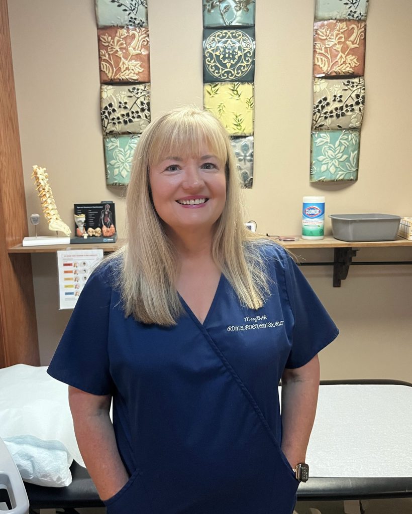
Mary Beth, Sonographer
Ultrasound imaging uses high-frequency sound waves that pass through your body. The sound waves are bounced off internal organs and tissues, and the waves are recorded and displayed by a computer. Ultrasound imaging works on the same principle as sonar, a technique to detect underwater objects used by both ships at sea looking for submarines and anglers looking for fish.
Ultrasound scanners usually consist of a computer, video screen, and a transducer. The transducer is a small, handheld device about the size of a bar of soap, attached to the scanner by a cord. The transducer generates the sound waves and detects them when they are reflected.
Most ultrasound exams are done by moving the transducer on your skin over the area to be examined.
Ultrasounds are also referred to as ultrasound scans, sonography, and sonograms. Some specific types of ultrasounds are abdominal, doppler, and echocardiogram.
Examples of Uses:
Ultrasound can be used to view, monitor, or diagnose
- abdominal organs; gallbladder, liver, kidneys, aorta
- the heart
- blood flow
- breast
- muscles and tendons
- soft tissue of any part of the body
- neck arteries (carotid)
- Prostate
- thyroid, including biopsies
Because ultrasound provides real-time images, it also can be used to guide procedures, such as a biopsy, in which a needle is used to obtain cells from an abnormal area for laboratory testing. This can be especially helpful in the guidance of pain relief injections. These are for low back pain, upper back pain, neck pain, jaw pain (TMJ), the wrists and the hands. All the areas you have pain… we can help!
Preparation: Wear comfortable, loose clothing. Additional preparation depends on the type of ultrasound exam you will have. Those ultrasounds done in the abdominal area should be fasting, 6-8 hours. Those done in the lower abdomen are done with a full bladder. Patients having their knees examined should wear shorts or pants that easily can be brought 4 inches up above the knee. At appointment scheduling, you will be advised of the specific prep for your exam.
During the Exam: You will be asked to lie on an exam table. The technologist will apply a water-based gel to the area being examined to help the transducer more easily move over the skin. The technologist will press the transducer firmly against the skin and move it back and forth to obtain an image of the area. Some patients require more pressure. Most ultrasound exams are quick, easy, and painless.
Time Required: 20 to 45 minutes.
Benefits:
- Ultrasound scanning is noninvasive
- Ultrasound uses no ionizing radiation
- Can be used on all patients regardless of barriers such as pacemakers
- Using real-time imaging makes ultrasound a good tool for guiding minimally invasive procedures such as needle biopsies and injections
Risks:
There are no known harmful effects for standard diagnostic ultrasound exams.
Sonographer:
Mary Beth, our sonographer, has nearly 30 years experience in diagnostic imaging. She has worked with Dr. Freeman since 1999. Mary Beth is board certified in Echocardiography, Vascular, Abdominal, OB-GYN, and Breast sonography. Additionally, holds certifications in Cat Scanning, Mammography and Radiologic Technologies. In 2013, Mary Beth became, and still to this day, the only Board Certified Sonographer in Musculoskeletal Ultrasound, in South Carolina.
In 2001, she was recognized and honored with a pioneer award becoming one of the first 500 in the country to be board certified in breast sonography and the first in South Carolina! About the same time, Dr. Freeman and Mary Beth began learning musculoskeletal ultrasound. Today, using this technique hundreds of our AMS patients have benefited from fast relief from musculoskeletal pain.